Industry content

Types of Dental X-Rays: What Your Patients Need to Know

Date Posted: 07.08.22

As a dental surgeon, dentist, or other healthcare professional providing dental services, you are focused on providing the best dental care possible to your patients. While keeping patients healthy is your number one priority, it’s important to realize that many — if not most — of your patients are in the dark about how dentistry works. This includes limited knowledge of all the different types of dental X-rays and imaging your practice provides or their care plan requires.

The following offers some examples of ways to explain different types of dental X-rays and imaging to your patients in layman’s terms. Whether ordering an X-ray, scan, or imaging study for screening or diagnostic purposes, it is always a good idea to know how to bypass the medical jargon and explain what X-rays are being taken and why. You should be able to communicate this in terms that will help your patients understand how these procedures fit into their overall plans of care. An informed patient will be more relaxed and more compliant with your recommended procedures and treatments, a win-win for both you as a significant healthcare provider and your patient as someone seeking compassionate care.

Intraoral vs. Extraoral Dental X-Rays

Patients understand that getting regular teeth X-rays is part of routine dental care. What they probably don’t know is why some (intraoral X-rays) are taken inside the mouth and others (extraoral X-rays) are taken outside of the mouth, what you are looking for with each procedure, and why you are choosing one procedure over another.

Before getting into the details about particular types of mouth X-rays or why you are ordering a specific study, it is a good idea to raise the patient’s awareness of the efficacy of undergoing routine screening tests and, if necessary, further diagnostic procedures. For instance, patients should be made aware that X-rays and other studies are helpful in:

  • Locating decay between teeth and beneath existing fillings
  • Indicating whether any abscesses are present
  • Showing any changes in the bone or root canal that indicate signs of infection
  • Determining whether there is any indication of jaw bone loss
  • Ruling out cysts and tumors
  • Determining the position and condition of teeth as you prep them for procedures like implants, braces, or dentures
  • Determining if any teeth are impacted

Types of Dental X-Rays

Patients often wonder why you’re recommending a bitewing vs. periapical X-ray dental procedure or why you selected a single tooth bitewing X-ray vs. full mouth. They might also want to understand why and when you require occlusal or panoramic X-ray vs. a cephalometric projection, what it means, and when you order a CBCT. The following should help explain the differences between these and other types of dental X-rays.

Intraoral Dental X-Rays

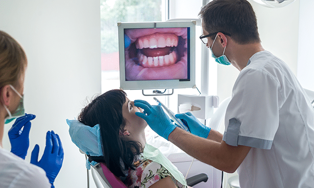
Intraoral Dental X-rays are the types of teeth X-rays your patients will be most familiar with. They are taken in the office while the patient sits in the chair. While patients may be familiar with the procedures for intraoral X-rays, chances are they don’t understand why you are taking them.

  • Bitewing X-rays. Practically everyone who visits a dentist has had bitewing X-rays taken. Patients are used to getting them during preventative maintenance visits and when they make an appointment to see you to relieve their tooth discomfort. Patients might not realize how important up-to-date bitewing X-rays are in allowing you to visualize decay that can exist between teeth or under gum lines, locate the source of tooth pain, detect changes in bone due to gum disease, and help determine the proper fit of a crown. The process of obtaining and reviewing bitewing X-rays has gotten a lot faster in recent years as the use of X-ray films has given way to a sensor that instantly relays images to a computer, eliminating the need to wait for X-ray films to develop.
  • Periapical X-rays. While bitewings are sufficient if only a portion of the tooth needs to be seen, there may be instances where periapical X-rays are required, such as when you suspect damage to the tooth root tip. If the patient is experiencing potential jawbone issues, a periapical X-ray enables you to see the whole tooth, from the top of the crown to the jaw area where the root is attached. In these instances, your patient should understand that periapical X-rays are a better choice as they allow you to see all the teeth in the relevant portion of the upper or lower jaw.
  • Occlusal X-ray. The occlusal X-ray provides important information for patients who present with issues regarding teeth that have not yet erupted, have extra teeth crowding their mouth, or present with a cleft palate or a fracture. With an occlusal X-ray, you can see inside the roof or the floor of the mouth to get a full visual of tooth development and placement. Because these X-rays can track the development and placement of an entire arch of teeth in either the upper or lower jaw, they not only help with diagnosing and treating these issues but also are invaluable in helping you locate cysts, abscesses, and growths.

Extraoral Dental X-rays and Imaging

There are several types of extraoral X-rays. While the most common type — the panoramic X-ray — can be part of a routine dental checkup or in preparation for a dental procedure, other types of extraoral diagnostic tools are needed to detect more severe dental problems in the jaw and skull.

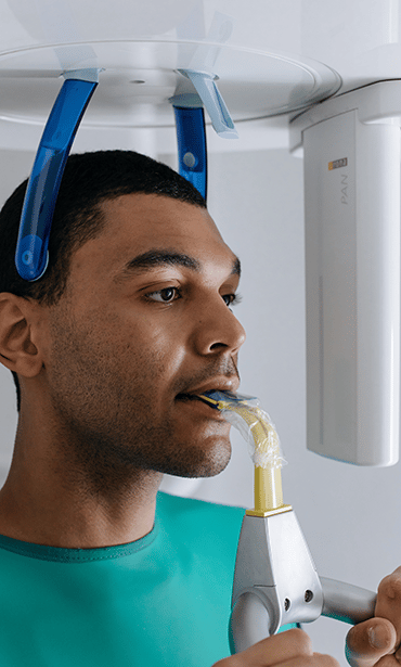
  • Panoramic X-ray. When undergoing a panoramic X-ray, the patient stands in a special machine that takes a single two-dimensional image of their upper and lower teeth. You may order a panoramic study for patients with a history of dental work to check that everything is holding up well or for patients about to undergo a significant procedure. Panoramic X-rays are also useful to diagnose serious issues like jaw tumors and cysts.
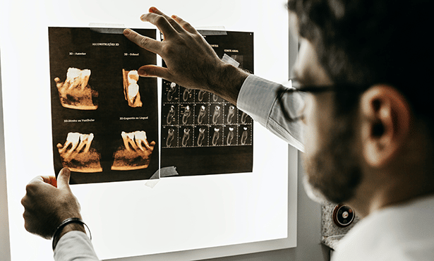
  • Tomograms. This type of X-ray is designed to allow you to examine mouth structures that are blocked from view using other X-ray methods. With a tomogram, a layer or slice of the mouth under investigation becomes accessible while other layers are blurred out.
  • Sialogram. You may order a sialogram for patients presenting with dry mouth, dry eyes, or where specific salivary gland problems are suspected. Your patient needs to understand that since salivary glands are soft tissue that are not rendered on an X-ray, this test requires a dye to be injected into the salivary glands to produce an image that will show up on the X-ray film.
  • Dental computed tomography. You may order a dental CT — an imaging method that allows you to see the interior of the mouth and face in 3D — to locate potential problems in the bones of the face. You might order this test if you are seeking information about facial fractures, cysts, or tumors.
  • Cephalometric projection. Orthodontists often have a patient undergo cephalometric projection — an X-ray of one side of the head — to gather information about a patient’s teeth and jawbones when formulating a treatment plan. A dentist may use cephalometric projection to see a patient’s throat structure clearly. This may be required if they suspect the presence of throat lumps or cancer or to determine the cause of sleep apnea.
  • Cone beam X-ray. Also called a CBCT, this imaging method uses computer technology to convert 2-dimensional pictures of a patient’s teeth and surrounding bone into a 3-dimensional image. The benefit of this procedure is that it allows you to see the entirety of the patient’s teeth and surrounding bones.
  • Digital imaging. With 2-D digital imaging, not only will you be able to see the images on your computer screen in mere seconds and share these images with colleagues, but you can also enhance and enlarge the images. This enables you to examine a tooth, for example, in a way that is impossible in an oral exam alone.


Enhance Patient Communication with Cloud 9 Software

Today’s patients are eager to become involved in their own care. With all of the information — and disinformation — about healthcare on the internet, you owe it to your patients and your practice to be clear and forthcoming about your patient’s treatment plan. Patients are privy to the information they need to make sound decisions about their care and demand superior service in all aspects of dental practice-patient communications. This means they want to know that — from the time they set their initial appointment to receiving their treatment plan and services to receiving their bill — they are working with a patient-centered practice with their best interests in mind.

Cloud 9 Software’s cloud-based dental practice management software provides the level of efficiency and transparency that orthodontics, pediatric dentistry, group practices, dental service organizations (DSOs), and orthodontic service organizations/OSOs need and that their patients require. As the leading provider of orthodontic cloud solutions, we can help you navigate opening a dental practice and managing it.

Our platform was designed to improve staff productivity, increase user efficiency, and minimize workflow stages — all important elements of a great five-star patient experience. Request a free demo today to learn more about how Cloud 9 can help you manage your practice.